CHAPTER 7
CARDIO – VASCULAR SYSTEM.
HISTORY OF THE STUDY OF BLOOD-CIRCULATION.
It is known that the blood is circulated in a closed heart-vascular system consisting of two laps of the heart and circulatory system.
In the evolution of the circulatory system are noted three areas:
1. Formation of the closed-loop system was carried out by development of a capillary system between arterial and venous areas bloodstream.
2. Differentiation specialized on the muscle layer of the heart.
3. The formation of two closed circles of circulation -large and small.
The opening laps of blood circulation associated with the name of the English scientist W. Harvey, who in 1628 "among the deep darkness and difficult imaginable now confusion that reigned in views on the activities of the animal and the human body,... spied one of the most important functions of the body - blood circulation - and thus laid the foundation of a new department of human knowledge precise - Animal Physiology» (Pavlov).
Until W. Harvey in science dominated presentation K. Galen according to which the blood is formed in the stomach from food, which arrives in the liver, followed by venous vessels delivered to the authorities, where consumed in the process of life. Harvey made a simple calculation: if the heart is for 1 hit throws 16 ml of blood (this is a wrong notion, in fact, stroke volume is 40-70 ml), then for 1 hour cardiac output makes 6720 ml. Assuming that the blood is spent authorities, the food consumption is not possible to compensate for. Ligating the veins and arteries, Harvey was convinced that the blood moving in opposite directions on them (the arteries - from the heart, through the veins - to the heart). So there was an idea of the continuous circulation of the blood. The opening of the capillaries by M. Malpighi in 1687 made the circulatory system structurally complete.
The first presentation of a pulmonary circulation was expressed by M. Servetus in the XVI century. He suggested that the blood in the lungs is cleared from the "soot" and oxygenated.
English clergyman S. Hal (1733), held its first direct measurement of blood pressure in the arteries and veins of various animals. Brothers Webers found an inhibitory effect on the heart of the vagus nerve, and the brothers Tsiona and I. Pavlov - the stimulating effect of the sympathetic nerve.
Improving methods of investigation have provided new information about the heart and circulatory system, which led to significant advances in the field of cardiology practice. Creating a heart-lung machine allowed to carry out operations on the "dry" heart, and eventually a heart transplant.
IMPORTANCE OF THE CARDIO VASCULAR SYSTEM.
GENERAL CIRCULATION SCHEME.
The basic meaning of the cardiovascular system is to supply blood for tissues and organs. The blood moves continuously through the vessels, which gives it the ability to perform all vital functions. The cardio-vascular system includes the heart and blood vessels - the blood and lymph. The blood vessels carrying blood from the heart to the organs, called arteries and blood vessels those bring blood to the heart - veins. In humans, as in other mammals, the vascular system is closed, and is represented by two circles of circulation: large and small. To maintain blood pressure between heartbeats in this system must have the elastic vessel walls. In addition, the need for blood supply to various organs is not only different, but always changing depending on the activity of supplying blood to the organs. Arterioles organs, hard functioning at the moment, are expanding, capillaries are open, increasing blood flow to these organs, while in resting organs narrowed arterioles, capillaries are closed, the blood flow is reduced. Thus it becomes necessary to the existence of a number of special control and regulate the intensity of the circulatory mechanisms.
Circles of the circulation. Cardio vascular system is a closed system. Continuous blood flow is moving in two circles circulation: large and small.
The systemic circulation provides delivery to all organs and tissues of oxygen and nutrients. It starts in the left ventricle of the aorta, through which arterial blood is oxygenated is introduced into an artery branching off from it and running all the organs. In the bodies there is the artery tree-like branching into smaller arterial vessels, which pass into the arterioles breaking into a network of capillaries that permeate the organs and tissues. In the capillaries of the arterial blood gives tissues oxygen, nutrients, saturated with carbon dioxide and metabolic products (metabolites) and converted into the venous blood. Capillary venues are going in, they are merging. form small, then the large veins organs exiting from them and carrying venous blood. Veins, which carry blood from the trunk and lower limbs fall into the inferior vena cava, from the head and upper limbs - in the superior vena cava. Both vena cava bring venous blood into the right atrium. Here ends the systemic circulation (Pic. 13).
Pulmonary circulation is used to remove carbon dioxide, and oxygen saturation. It begins in the right ventricle of his carbonated venous blood to the pulmonary trunk, which in the lung is divided into left and right pulmonary arteries, passing into the capillaries. A dense network of capillaries entwines lung bubbles - the alveolus in the blood give them carbon dioxide, oxygen saturation, becomes bright red, and turns into the arterial blood. Arterial blood from the lungs back to the heart via the pulmonary veins 4 (2 from each lung) into the left atrium. From the left atrium arterial blood through the atria-ventricular orifice enters the left ventricle, from where the systemic circulation. Consequently, in the pulmonary arteries of the pulmonary venous flows, oxygen-poor blood, and the pulmonary veins - arterial blood. Thus, each portion of the blood after passing the pulmonary circulation, comes in a big circle and moves continuously along a closed circulatory system. The speed of blood circulation in a large circle is 22 seconds, in a small - 4-5 seconds (Pic. 13).
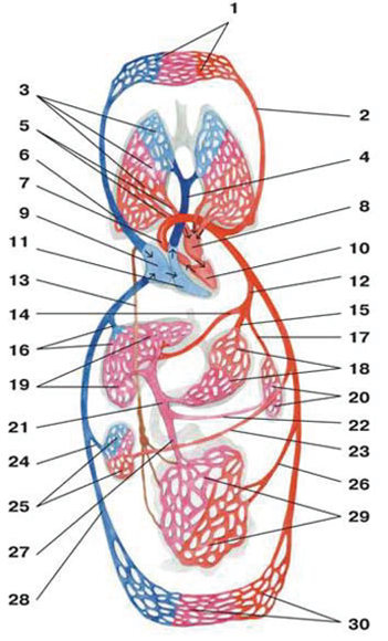
Рис. 13. Схема большого и малого кругов кровообращения
1 — капилляры головы, верхних отделов туловища и верхних конечностей; 2 — левая общая сонная артерия; 3 — капилляры легких; 4 — легочный ствол; 5 — легочные вены;
6 — верхняя полая вена; 7 — аорта; 8 — левое предсердие; 9 — правое предсердие;
10 — левый желудочек; 11 — правый желудочек; 12 — чревный ствол; 13 — лимфатический грудной проток; 14 — общая печеночная артерия; 15 — левая желудочная артерия; 16 — печеночные вены; 17 — селезеночная артерия; 18 — капилляры желудка; 19 — капилляры печени; 20 — капилляры селезенки; 21 — воротная вена; 22 — селезеночная вена; 23 — почечная артерия; 24 — почечная вена; 25 — капилляры почки; 26 — брыжеечная артерия; 27 — брыжеечная вена; 28 — нижняя полая вена; 29 — капилляры кишечника; 30 — капилляры нижних отделов туловища и нижних конечностей.